Diagnosezentrum Donaustadt. All Health Insurances & Private.
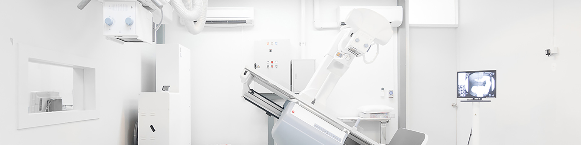
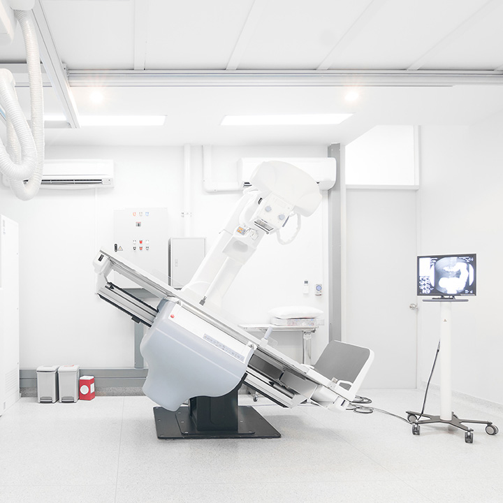
We have an ultra-modern, fully digital fluoroscopy system that works completely without film. As a result, the shortest possible examination times and the lowest possible radiation dose are standard for you at our center.
Please note the separate information on stomach and duodenum X-ray on this page.
Alth & Partner Fachärzte für Radiologie GmbH
Your attending physician has referred you for an X-ray examination of the stomach and duodenum. So that we can assess your stomach and duodenum as well as possible, please follow these preparation recommendations:
Please do not eat or drink anything for 4 hours before the examination so that your stomach and duodenum are completely empty. Unfortunately, this is necessary so that we can examine your stomach and duodenum. Apart from this, no further preparation is needed.
First, you will stand on the examination table in the X-ray examination room. You will be given about 1 tablespoon of effervescent powder to swallow, followed by about the same amount of water. This produces gas in your stomach, which distends it. This is necessary so that the stomach can be clearly imaged and examined.
Please do not burp up the air that forms in your stomach.
Next, you will be given a barium-containing X-ray contrast agent – it does not taste very pleasant, but unfortunately it is necessary for the examination.
After you have drunk the contrast agent, the examination table will bring you into a lying position on your back. Please then turn once all the way around your body axis so that the X-ray contrast agent can coat all parts of the stomach.
Several images of your stomach and duodenum are then taken in different positions. Finally, images are taken while you are standing – then the examination is complete. The examination is painless and safe.
Kidney X-ray (intravenous pyelography) is used to detect changes in your kidneys, ureters, and bladder. This examination requires the administration of an iodine-based contrast agent into a vein.
So that we can properly assess your kidneys, ureters, and bladder, please follow these preparation recommendations:
2 days before the examination:
Low-residue diet: plenty of fluids, no fruit, no vegetables, no legumes, no whole-grain products, no fatty foods, no sweets.
1 day before the examination:
Lunch: low-residue diet (as above)
Evening: a small low-residue meal, plenty of fluids.
For breakfast, we recommend tea, coffee, and, if desired, some rusks – plenty of fluids are allowed.
You should not eat anything until the time of the examination, but you may continue to drink without any problem.
You lie on the X-ray examination table.
First, an X-ray image is taken without contrast agent to detect kidney stones. Then an iodine-based contrast agent is administered through a vein in your arm.
The contrast agent is carried by the bloodstream to the kidneys and excreted by the kidneys through the urinary tract.
This process is examined with several X-ray images, which allows us to detect changes in your kidneys, ureters, and bladder.
The examination usually takes less than 30 minutes and is painless apart from the small needle prick for the contrast agent injection.
To rule out a possible intolerance to the contrast agent, please fill out the questionnaire.
You should not eat anything until the time of the examination, but you may drink without any problem.
First, you will be given a warm foot bath for the affected leg in the examination cubicle – this serves to dilate the veins on the top of the foot.
You will then be asked into the examination room, where, while you are in a semi-reclining position, the doctor punctures a vein on the top of your foot. This is not always easy, so a second puncture is sometimes necessary to ensure that the needle is positioned correctly and securely in the vein.
Next, an iodine-based contrast agent is administered into the vein on the top of your foot through an extension tube, making the veins of the leg visible.
Several images are taken with the leg rotated into different positions.
Finally, you will be asked to bear down with your abdomen as if having a bowel movement. This increases the pressure in the abdomen, stops the blood flow in the veins, and makes any backflow through diseased venous valves visible.
X-ray examinations and bone density measurements are performed immediately – without prior booking – between 7:00 AM and 6:00 PM. For all other examinations (CT / Computed Tomography, MRI / Magnetic Resonance Imaging, Mammography, Ultrasound / Sonography, Fully Digital Fluoroscopy, X-ray Pain Therapy, IVP / Kidney X-ray), please book an appointment by phone.
We respect your privacy. Cookies and third-party plugins are only used with your consent, and our website is ad-free. For more details, please see Privacy Policy.
X-ray examinations and bone density measurements are performed immediately – without prior booking – between 7:00 AM and 6:00 PM.
Mammography appointments can now be booked online:
For all other examinations (CT / Computed Tomography, MRI / Magnetic Resonance Imaging, Ultrasound / Sonography, fully digital fluoroscopy, X-ray pain therapy, IVP / kidney X-ray), please book an appointment by phone.
